
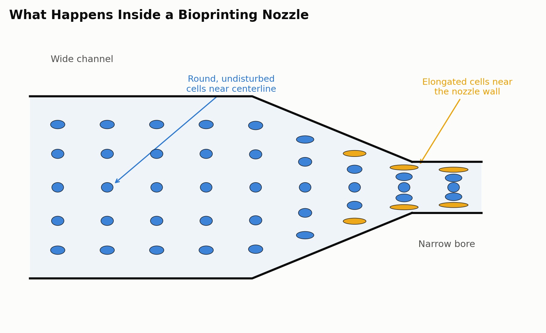
 The same narrowing that sharpens a printed strand also narrows the channel the cells have to survive passing through.
A bioprinter is the only additive process whose feedstock is already alive, and that one fact sets a floor under its resolution that no amount of mechanical refinement has moved. A typical mammalian cell runs 10 to 20 micrometers across. An extrusion bioprinter’s nozzle bore runs 100 to 500 micrometers, and the bead it lays down measures 250 to 600 micrometers wide in most published work (Guida et al., 2024). Even a finely tuned extrusion system is printing strands tens of cells wide, not placing individual cells. The resolution problem starts before the printer ever turns on, in the simple mismatch between the size of the thing being deposited and the size of the channel doing the depositing.
Shrinking the nozzle looks like the obvious fix, and it runs into a different limit almost immediately. Below roughly 100 micrometers, cell-laden bioinks start clogging the orifice outright, since cells and cell clusters are now a meaningful fraction of the channel’s own diameter (Jentsch et al., 2021). Push past that limit anyway and a second effect arrives before the first one even clears: shrinking the bore raises the shear rate at the wall for the same flow rate, the way narrowing a pipe speeds up the water moving through it. Fluid dragged past a stationary wall decelerates sharply right at that boundary, and the cells riding closest to the wall see the steepest velocity gradient in the channel. Membranes stretch, deform, and past a threshold the cell either survives compromised or doesn’t survive at all. Across the published range, extrusion viability runs anywhere from 40 to 95 percent, swinging on exactly this tradeoff between applied pressure, nozzle diameter, and bioink viscosity (Cytion, 2023). Finer resolution and cell survival pull on the same rope from opposite ends, because both are set by the same wall.
Light-based printing sidesteps the mechanism rather than fighting it. Vat photopolymerization cures a bioink resin layer by layer with a projected light pattern, so there is no bore for cells to be pushed through and no wall for a velocity gradient to form against. Remove the nozzle and the dominant source of shear stress goes with it. Commercial digital light processing systems now resolve features down to 10 to 35 micrometers (CELLINK, 2025), and a 2024 review puts stereolithography and DLP variants in an 8 to 10 micrometer range, overlapping the lower end of single-cell scale for the first time (Guida et al., 2024).
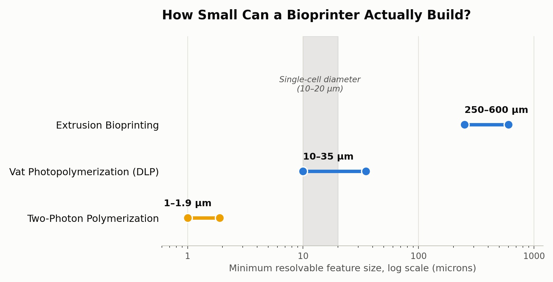 Extrusion deposits well above single-cell scale. Vat photopolymerization is the first method to reach it, and two-photon polymerization overshoots it by an order of magnitude.
Push the same mechanism further and two-photon polymerization cures a voxel at the focal point of two overlapping infrared beams, with no mask or projected pattern spreading the exposure, and resolves features down to 1 to 1.9 micrometers, smaller than the cell it is meant to pattern around (Guida et al., 2024). That precision buys almost nothing for tissue engineering at useful scale, though, because point-by-point laser scanning confines practical part size to well under a millimeter. A process fine enough to pattern a single organelle cannot build a construct a surgeon could hold.
Acoustic droplet ejection splits the difference, keeping inkjet’s drop-on-demand logic while removing the nozzle wall inkjet still relies on. A focused acoustic pulse ejects a droplet straight from an open reservoir surface, no channel involved. Simulated peak shear stress comes out at roughly a third of what a conventional 150 micrometer microvalve nozzle produces for the same task, and more than 90 percent of cells survive the ejection with no measurable change to stem cell morphology or differentiation capacity afterward (Jentsch et al., 2021). Resolution and cell survival stop competing exactly where the nozzle wall disappears.
None of these approaches erase the constraint, they just move it. Vat photopolymerization needs bioinks formulated around a photoinitiator, which narrows the materials palette compared to anything a syringe can simply push through. Two-photon polymerization trades resolution for build volume outright. Acoustic ejection needs droplet-forming rheology, which rules out the high-viscosity, cell-dense pastes extrusion handles without complaint. The nozzle wall was never the only constraint, just the most visible one, and removing it reveals whichever constraint was standing behind it. The real determinant of bioprinted tissue resolution was never the hardware. It is the cell, at 10 to 20 micrometers, setting the scale that every printing method is ultimately measured against.