
The first extrusion-printed tissue constructs, built in the early 2010s, could hold a shape and keep cells alive near the surface. What they couldn’t do was feed themselves. Anything thicker than about 200 micrometers from an open edge starved for oxygen and nutrients within days, because diffusion alone can’t reach cells buried deeper than that without a real circulatory system to carry supply in. Bioprinting had solved geometry. It hadn’t solved plumbing, and for a long stretch that gap defined the field’s ceiling.
Early workarounds treated vascularization as something to invite rather than build. Researchers seeded scaffolds with growth factors and left the body’s own vessels to grow in after implantation, a strategy that works for thin or highly perfused tissue but leaves anything larger, a section of liver or a patch of cardiac muscle, dead at the core before host vessels ever arrive. A 2022 review of vascularization strategies traces the field’s slow shift away from that passive approach, toward scaffolds engineered with their own channel networks meant to anastomose directly with a patient’s circulation rather than wait on it. That shift mattered because it reframed the problem: instead of asking a scaffold to attract blood vessels, printing was asked to fabricate the vessels themselves, as part of the tissue and on the same pass.
Getting there required accepting a tradeoff nobody loved in the printer’s own nozzle. Extrusion bioprinting pushes living cells suspended in hydrogel through a narrow channel, and the shear forces along that channel wall can shred cell membranes before the material ever reaches the build plate. An overview of induced shear stress in extrusion bioprinting put this plainly: viability in early extrusion work sometimes fell as low as 40 percent, a number that made the technique look like a poor substitute for gentler, lower-throughput methods like inkjet or laser-assisted printing. The fix turned out to be less about avoiding shear and more about respecting its limits. Keeping stress under roughly 5 kilopascals preserves survival above 90 percent for common cell lines, and a related line of work on shear stress preconditioning found that briefly exposing cells to controlled shear before printing actually toughens them against the shear that follows, turning what had been treated purely as damage into something closer to a manageable dose.
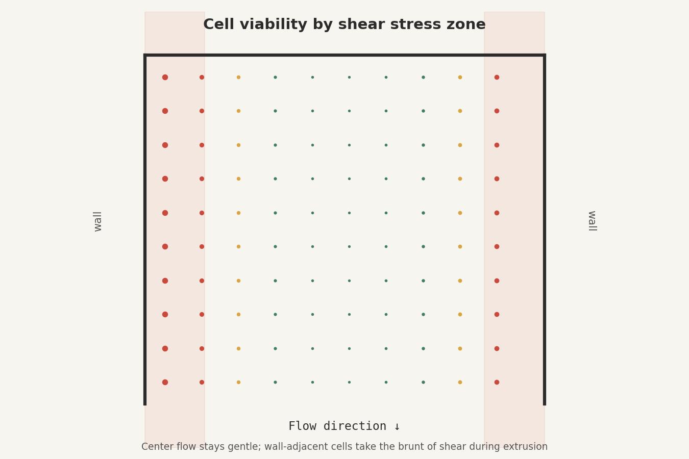 Keeping wall shear stress under roughly 5 kPa during extrusion is what separates a filament of living cells from a filament of dead ones.
Those two threads, better scaffold vasculature and gentler extrusion, converged this year in constructs that finally look like real tissue rather than a proof of concept. A 2025 study in Advanced Functional Materials describes thick adipose tissue printed with an integrated, branching vascular hierarchy built directly into the construct rather than bolted on afterward, a design that earlier scaffolds simply didn’t attempt because the fabrication tools weren’t there yet. That’s the pattern worth noticing: each generation of bioprinting didn’t discard the last one’s assumptions so much as inherit its constraints and design around them more precisely.
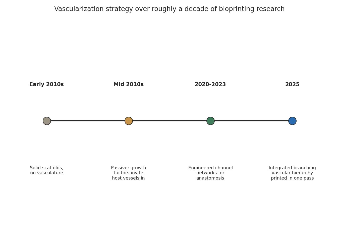 Vascularization strategy has moved from waiting on the host’s own vessels to fabricating the circulatory network as part of the print itself.
None of this makes lab-grown organs imminent. Thick, metabolically demanding tissue like liver or kidney still asks more of a printed vascular network than current resolution and perfusion rates can reliably deliver, and getting a construct to survive on a bench is a different problem from getting it to integrate into a living patient. But the trajectory is legible enough to take seriously. What began as a diffusion problem nobody had a good answer for has become an engineering problem with a widening set of partial solutions, and it’s the accumulation of those solutions, not one single breakthrough, that’s likely to decide how far bioprinted tissue actually gets.